Enquire Now
Intensity Modulated Radiation Therapy (IMRT)
IMRT addresses the shortcomings of 3DCRT and improves dose distributions.
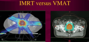
- IMRT is a computer-generated plan which uses multiple small fields called beamlets to generate complex field shapes to avoid critical normal structures.
- IMRT modulates a number of fields as well as intensity within each field, thereby ensuring greater control in delivering dose to the tumor while minimizing dose to normal structures.
- IMRT also helps with dose escalation to the tumor, thereby increasing the possibility of an improved treatment outcome.
- The decrease in toxicity also helps to improve the quality of life.


Treatment is carefully planned by using 3-D computed tomography (CT) images of the patient in conjunction with computerized dose calculations to determine the dose intensity pattern that will best conform to the tumor shape. Typically, combinations of several intensity-modulated fields coming from different beam directions produce a custom tailored radiation dose that maximizes the tumor dose while also minimizing the dose to adjacent normal tissues.
Because the ratio of normal tissue dose to tumor dose is reduced to a minimum with the IMRT approach, higher and more effective radiation doses can safely be delivered to tumors with fewer side effects compared with conventional radiotherapy techniques. IMRT also has the potential to reduce treatment toxicity, even when doses are not increased. IMRT delivers a low dose to larger volumes of normal tissue than conventional radiotherapy.
Currently, IMRT is being used most extensively to treat cancer. All types of tumors, e.g., of the prostate, head and neck, and central nervous system. IMRT has also been used in limited situations to treat breast, thyroid, and lung cancers, as well as gastrointestinal and gynecologic malignancies and certain types of sarcomas. IMRT may also be beneficial for treating pediatric malignancies.
Radiation therapy, including IMRT, stops cancer cells from dividing and growing, thus slowing or stopping tumor growth. In many cases, radiation therapy is capable of killing all of the cancer cells, thus shrinking or eliminating tumors.
There are several ways IMRT differs from conventional radiation therapy:
Employs powerful, advanced software to plan a precise dose of radiation based on tumor size, shape, and location.
Delivers radiation in sculpted doses that match the exact 3D geometrical shape of the tumor, including concave and complex shapes.
Adjusts the intensity of radiation beams across the treatment area as needed for laser accuracy.
Who will be involved in this procedure?
Most facilities rely on a specially trained team for IMRT delivery. This team includes the radiation oncologist, medical radiation physicist, dosimetrist, therapist, and radiation therapy nurse.
The radiation oncologist, a specially trained physician who heads the treatment team, sets an individualized course of treatment with the help of the radiation physicist, who ensures the linear accelerator delivers the precise radiation dose and that computerized dose calculations are accurate.
A highly trained radiation therapist positions the patient on the treatment table and operates the machine. The radiation therapy nurse provides the patient with information about the treatment and possible adverse reactions, as well as help in managing any reactions.
What equipment is used?
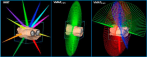
A medical linear accelerator generates the photons, or x-rays, used in IMRT. The patient lies on the treatment table while the linear accelerator delivers multiple beams of radiation to the tumor from various directions. The intensity of each beam’s radiation dose is dynamically varied according to the treatment plan.
Who operates the equipment?
The Radiotherapy Technologist operates the equipment from a radiation-protected area nearby. The RTT is able to communicate with the patient throughout the procedure. The therapist observes the patient on closed circuit television.
Is there any special preparation needed for the procedure?
Before planning treatment, a physical examination and medical history review will be conducted. Next, there is a treatment simulation session, which includes CT scanning, from which the radiation oncologist delineates the target volumes and normal organs. In some cases, a treatment preparation session may be necessary to mold a special device that will help the patient maintain an exact treatment position. The medical physicist uses the CT information to design the IMRT beams used for treatment. Several additional scanning procedures, including positron emission tomography (PET) and magnetic resonance imaging (MRI), might also be required for IMRT planning. These diagnostic images can be merged with the planning CT and help the radiation oncologist determine the precise location of the tumor target. It is necessary to insert radio dense markers into the target for more accurate positioning. Typically, IMRT sessions begin about a week after simulation. Prior to treatment, the patient’s skin may be marked or tattooed with colored ink to help align and target the equipment.
How is the procedure performed?
IMRT often requires multiple or fractionated treatment sessions. Several factors come into play when determining the total number of IMRT sessions and radiation dose. The oncologist considers the type, location, and size of the malignant tumor, doses to critical normal structures, as well as the patient’s health. Typically, patients are scheduled for IMRT sessions five days a week, as advised by their radiation oncologist.
At the beginning of the treatment session, the therapist positions the patient on the treatment table, guided by the marks on the skin defining the treatment area. If molded devices are made, they will be used to help the patient maintain the proper position. The patient may be repositioned during the procedure. Imaging systems on the treatment machine may be used to check positioning and marker location. Treatment sessions usually take between 10 and 30 minutes.
What will I feel during and after this procedure?
IMRT is painless. You will not feel or sense anything out of the ordinary during treatment. However, the machine can be stopped if you become uncomfortable. As treatment progresses, some patients may experience treatment-related side effects. The nature of the side effects depend on the normal tissue structures being irradiated. The radiation oncologist and the nurse will discuss and try to help you with any side effects.
Side effects of radiation treatment include problems that occur as a result of the treatment itself as well as from radiation damage to healthy cells in the treatment area. The number and severity of side effects you experience will depend on the type of radiation and dosage you receive and the part of your body being treated. You should talk to your doctor and nurse about any side effects you experience so they can help you manage them.
Radiation therapy can cause early and late side effects. Early side effects occur during or immediately after treatment and are typically gone within a few weeks. Common early side effects of radiation therapy include tiredness or fatigue and skin problems. Skin in the treatment area may become more sensitive, red, irritated, or swollen. Other skin changes include dryness, itching, peeling and blistering.
Depending on the area being treated, other early side effects may include:
- Hair loss in the treatment area
- Mouth problems and difficulty swallowing
- Eating and digestion problems
- Diarrhea
- Nausea and vomiting
- Headaches
- Soreness and swelling in the treatment area
- Urinary and bladder changes
There is a slight risk of developing cancer from radiation therapy. Following radiation treatment for cancer, you should be checked on a regular basis by your radiation oncologist for recurring and new cancers.
A state-of-the-art external radiation delivery system that uses advanced computer technology to create a three-dimensional model of a tumor and directs precisely-focused beams of radiation at the tumor with laser accuracy. By precisely modulating (controlling) the intensity of the radiation beams to conform to the tumor shape, IMRT can treat difficult-to-reach tumors with new levels of accuracy


