Oral cancer is one of the most common kinds of cancer in India, affecting a huge population. About 60-80% of the patients suffering from oral cancer are diagnosed in advanced stages, which contributes to a high mortality rate. Early detection of oral cancer is the most effective way to reduce the morbidity and mortality rate of oral cancer.
Persistent red and/or white patches, non-healing ulcers, progressive swelling or enlargement, unusual surface changes, sudden tooth mobility without apparent cause, unusual oral bleeding or epistaxis, and prolonged hoarseness are a few of the early signs and symptoms that are helpful in diagnosing oral cancer. These changes can be seen easily and are clinically detected through careful visual inspection and palpation of oral mucosa. Early evaluation of localized oral cancers which has not spread to surrounding tissue can be effectively treated, resulting in a more than 80 percent five-year survival rate.
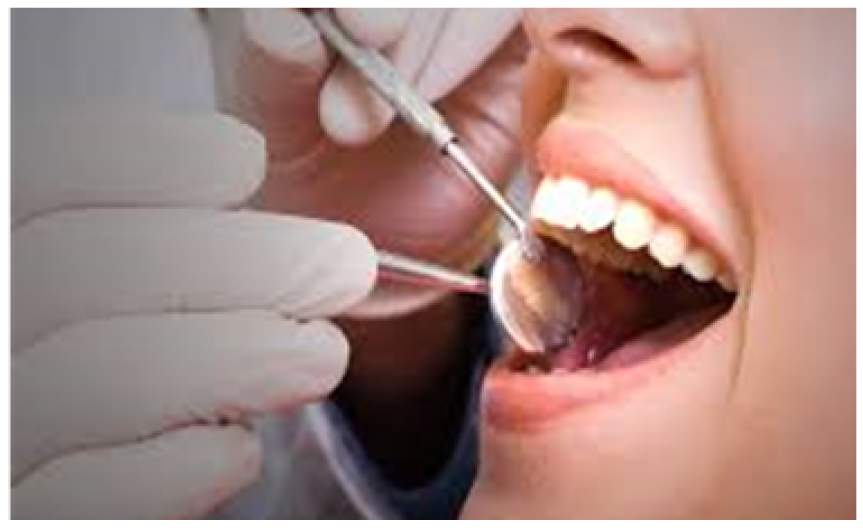
Visual examination, excision biopsy, brush biopsy, toluidine blue staining, light-based detection systems, biomarkers, DNA analysis, and laser capture microdissection are the various modalities used for oral cancer detection.
Toluidine blue staining is claimed to be a simple, inexpensive, and sensitive adjunct tool for identifying early oral cancer. Its application is very easy and fast – Toluidine blue dye is applied on the area of the suspected lesion for 30 sec and then the staining pattern is evaluated
Autofluorescence imaging contributes to diagnosis and helps to discern lesions that warrant a biopsy. It is based on the concept that dysplasia or cancer causes changes in the fluorescence of the mucosa. The condition produces a decrease of green fluorescence, which darkens the mucosa, whereas normal mucosa has light-green fluorescence.
Many chemical and inorganic compounds, proteins, peptides, and electrolytes are found in saliva. Molecular studies have identified more than 100 biomarkers in human saliva that acts as indicators of pathological processes and carcinogenesis, such as viruses, cytokines (IL-1b, IL-8, TNF-α), protein receptors (CD44), DNA and RNA markers that are overexpressed in a carcinogenic process. Diagnosing saliva using nanotechnology and molecular technologies to detect oral cancer is non-invasive and cost-effective. The disadvantage of this method is most people have a small amount of the markers that prevent detection, the levels of these markers tend to increase when the disease progress. Some cancer patients may never have high-level markers and even in the presence of elevated levels, they do not always indicate cancer, as they may be related to other disorders.
DNA image cytometry determines the malignant potential of cells by measuring ploidy status. A computer-assisted technique was recently developed to detect variations in cellular DNA content. Cancer growth is aided by genomic instability, and aberrant DNA content may differentiate dysplastic tumours that may lead to cancer. DNA ploidy analysis as an adjunct to conventional cytology assessment of cytobrush samples is useful for the detection of oral cancer.
Laser capture microdissection provides an ideal method for the extraction of cells from specimens in which the exact morphology of both the captured cells and the surrounding tissue are preserved.Rapid immunohistochemical staining methods paired with LCM allow for more precise microdissection of cellular subsets. LCM may also be utilised to identify biomarkers and build protein fingerprint models for early diagnosis of oral squamous cell cancer.
Brush biopsies are painless, chair-side tests that may be performed to analyse any suspicious lesion, including typical tiny white and red oral lesions to rule out dysplasia. Cells are collected from the whole thickness of the oral epithelium during the brush biopsy. Tissue samplings by scalpel biopsy and subsequent histological examination have been the cornerstone for diagnosing premalignant and malignant oral diseases.

Written by