Enquire Now
PET CT
What is PET?
The advantages of doing a PET are
High sensitivity: PET scans can pick up cancer sites that other imaging tests like CT or MRI sometimes cannot. Differentiating tumor from fibrosis: Some cancer treatments, especially radiation therapy, can cause tumor areas to fibrose. Conventional imaging techniques cannot differentiate tumor from fibrosis, whereas PET can.
Evaluating treatment effectiveness: Change in a tumor’s metabolic activity occurs much before change in its size and hence can more effectively demonstrate response to treatment. PET enables doctors to see these changes better than other imaging methods. Also, patients shown to be responding to treatment on PET have a much better prognosis than those who don’t.
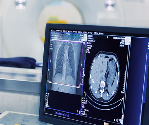
Why is CT done along with PET?
In addition to PET, the latest machines have a built-in CT scanner. CT scanners emit a series of very narrow beams that travel through the body in an arc-shaped path, unlike an X-ray machine, which uses just a single beam. As a result, CT scanners produce a final picture far more detailed than basic X-ray machines.
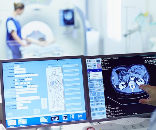
The addition of CT scanner has the following advantages:
- It helps make the PET images more accurate
- The PET images can be superimposed on to the CT images, so doctors can localize the site of PET abnormality correctly.
- Fusion of PET and CT images gives more information about tumor characteristics and metabolism.
Can PET/CT detect all tumors?
The ability to detect the tumors depends on their size, metabolic characteristics, and location. PET can detect tumors of 1 cm or more with very high accuracy. Tumors between 5mm and 1cm fall in the gray zone and can sometimes be missed. Tumors smaller than 5 mm and microscopic disease cannot be picked up on PET or in other imaging procedures, for that matter.
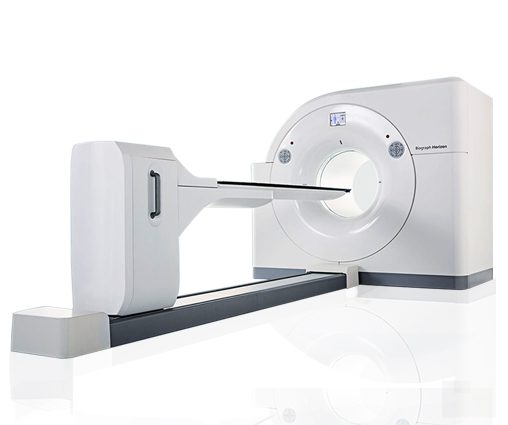
What is Open Gantry PET/CT?
The Open View gantry design improves patient comfort by reducing the claustrophobic effect associated with a long tunnel and also allowing for easier patient access.
What is 4D PET?
4D PET technology, which is available at the Mahatma Gandhi Cancer Hospital & Research Institute, is a sophisticated technique of acquiring a PET scan that compensates for organ motion during breathing, which can make images of abnormalities look hazy. For cancer patients, this advanced technique helps doctors visualize tumors, especially in the lungs, better and guide radiation therapy far more precisely than without it, allowing them to target the treatment only to the tumor itself and not surrounding healthy tissue.
What are the required preparations?
- The patient should be fasting 4-6 hours before the scan. Drinking water is allowed.
- If you are diabetic, kindly inform us before the scan. The patient can take anti diabetic tablets 4-6 hours prior to the scheduled time for the scan, along with a light breakfast.
- If patients are on IV fluids don’t use DNS 6 hours before study.
- Your appointment may be rescheduled/canceled in case of high blood glucose levels.
- Patients are requested to remove all the ornaments before coming for the scan.
- Patients are instructed to bring discharge summary, and all their previous investigation reports (X-Ray, Ultrasound, CT Scan CD/Films or MRI Scan CD/Films) at the time of scan.
What happens to the radioactivity that is injected?
After emitting the radiation, these elements convert into more stable forms. This process of conversion from a radioactive state to a stable state is called decay. Radioactive elements decay naturally, becoming less and less radioactive as time passes. The rate at which they decay is measured in terms of half-life. One half-life is the time required for half of the radioactive material to decay. After 4 half-lives, approximately 95% of the activity is decayed. And your body excretes some of the radioactive materials, too, so the amount of radiation decreases with time.
What are side effects of PET/CT scan?
Side-effects to nuclear medicine agents are extremely rare. The amount of radioactive substance injected is an incredibly tiny fraction of a gram. Very rarely, side-effects can occur either from the organ specific agent or preparation agents used, but they generally occur less than once in every 50,000 cases.
Can I Have a PET scan even if I do not have cancer?
A PET scan can also be done for conditions other than cancer, such as:
- Unexplained fever
- Inflammatory diseases such as arthritis
- Chronic infective diseases
- Brain disorders such as dementia, memory loss, movement disorders, or epilepsy
- Cardiac abnormalities.
Center of Excellence in Time of Flight PET-CT Scan
- FDG PET-CT Scan
- PSMA PET-CT for Prostate Cancer
- Ga-DOTANOC Scan for Neuro Endocrine tumors
- Metabolic Imaging for Epilepsy, Dementia, Parkinsonism
- Metabolic Biopsy
- Cardiac PET-CT for Viability & Inflammation
- F-DOPA PET/CT
- 68 Ga - FAPI PET/CT
- Choline PET/CT
Salient Features
- Revolutionary unique Time of Flight (TOF) technology
- LSO Crystal Technology
- Best Image Quality
- Low Radiation dose to the Patient
- Faster Image Acquisition
- Pediatric PET-CT with Low Dose Protocols
- Better image quality in Obese Patients
- Artificial Intelligence in Neuro Imaging