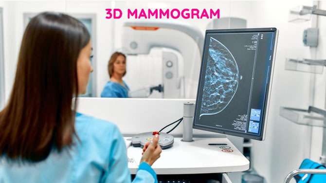
A 3D Mammogram is an imaging test in which multiple images of the breast are taken to create its 3-dimensional image. It is used as a screening modality in asymptomatic women for detecting possible breast cancer. It can also be used to evaluate women having symptoms such as nipple discharge or lump or pain in the breast.
If breast cancer is detected early when it is small and has not spread to other parts, the chances of successful treatment are more.
There are no fixed guidelines for breast cancer screening in India. American Cancer Society recommends- those between 40 and 44 years should have the option of getting it done, yearly Mammograms between 45 and 54 years, and once in 2 years after that. In people who have a high risk of breast cancer (who have a history of breast cancer in the family members, gene mutations, or history of radiation to the chest at a young age), screening should start early, between 25-30 yrs.
3D Mammograms can detect breast cancer early and in a greater number of asymptomatic women when compared to other modalities. It reduces the number of false positives (False positive means the test result is suggestive of cancer but in reality, there is no cancer).
This procedure is somewhat similar to a CT scan of the breast, but with a much lesser and a fraction of radiation. A standard 3D Mammogram screening includes a 2D Mammogram along with a 3D Mammogram and this combination improves the possibility of picking up cancer early.
Advantages of a 3D Mammogram in combination with a 2D Mammogram are:
- Possibility of detecting early cancers in a greater number of people
- Reducing false positives and requirement of additional imaging
- Improving image clarity in dense breasts which are generally difficult to screen as the disease is camouflaged by the denseness of the breast tissues.
However, there is slightly more radiation compared to a standard Mammogram, but the benefits outweigh this. As with any test, it may also miss some very early cancers.
The procedure is simple, a clear plate is used to compress the breast, and images are taken from multiple directions. All these images are processed by the computer and produce good quality images for interpretation by the radiologist.
Additional imaging like ultrasound, MRI, or biopsy may be required depending on the result of a 3D Mammogram.
It is always advisable to carry all the previous images and reports for comparison and better interpretation.

Written by
Dr. Madhuri Palli,
Senior Radiologist,
Mahatma Gandhi Cancer Hospital and Research Institute, Visakhapatnam